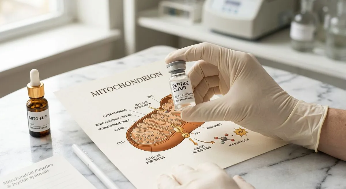
Humanin and its analogues (HNG, S14G-humanin) are research peptides. They are not approved for human administration by any regulatory authority. All information on this page is provided strictly for scientific and educational purposes. UNIK LAB does not provide medical advice.
Most people working in longevity research know about NAD+, senolytics, and GLP-1 agonists. Far fewer know about humanin. That's been true since 2001 — when a Keio University team published a paper in PNASdescribing a 21-amino acid peptide encoded not in the nuclear genome, but in mitochondrial DNA. They called it humanin because the only library that yielded the peptide's neuroprotective signal was from the surviving neurons of an Alzheimer's disease patient's brain. The intact cells. The ones that hadn't died.
That discovery has been quietly accumulating research significance for over two decades. Humanin circulates in human blood. Its levels decline with age. Higher levels correlate with longevity in population studies. It signals through a receptor (FPRL2) expressed in neurons, immune cells, and multiple peripheral tissues. And its analogue — S14G-humanin (HNG) — shows approximately 1,000-fold higher potency than the natural sequence in some bioassays.
1. Discovery: The Mitochondrial Genome Encodes Bioactive Peptides
Hashimoto Y, Niikura T, Tajima H, Yasukawa T et al. (2001, PMID 11371646, PNAS) disrupted the assumption that mtDNA encoded only 13 proteins. Working from an expression library derived from surviving neurons in an Alzheimer's disease brain, they identified a small open reading frame within the 16S ribosomal RNA gene of human mtDNA that, when expressed, protected neurons from Alzheimer's-associated death signals including Aβ peptides, mutant presenilin-2, and V642I APP.
This discovery opened the field of mitochondrial-derived peptides (MDPs). Humanin was the first. MOTS-c (published 2015) was the second. Researchers now believe there may be many more.
2. Humanin Structure and Variants: HN, HNG, S14G
- Natural humanin (HN): 21 amino acids (core sequence), MW ~2,485 Da, encoded in the 16S rRNA region of human mtDNA
- S14G-humanin (HNG): Single substitution at position 14 (serine → glycine) producing ~1,000-fold higher potency in neuroprotection assays. HNG is the preferred research tool for studies requiring practical working concentrations.
- Structural note: Humanin contains a disulfide bond (via the cysteine residue) important for its three-dimensional structure and bioactivity. Research protocols must account for oxidation-reduction conditions.
3. Receptor Biology: FPRL2, gp130Rβ, and the Tripartite Receptor Complex
FPRL2 (Formyl Peptide Receptor Like 2) / FPR2: The primary high-affinity receptor for humanin. A GPCR expressed on neurons, immune cells, and several peripheral tissues. Activates PI3K/Akt, ERK1/2, and STAT3 signalling.
gp130Rβ — Tripartite cytokine receptor complex: Composed of gp130, CNTFR, and WSX-1. Structurally similar to receptor systems for CNTF and IL-6. Mediates the JAK/STAT3 signalling arm of humanin's neuroprotective activity.
Intracellular IGFBP-3 interaction: Humanin also interacts with IGFBP-3 intracellularly, proposed to mediate some anti-apoptotic effects by modulating IGFBP-3's pro-apoptotic nuclear activity.
4. Neuroprotection: The Core Research Signal
Zárate et al. (2019, PMID 31214013, Front Aging Neurosci) demonstrated a key dimension: humanin is not just protective when administered exogenously — it is produced and secreted by astrocytes, and this astrocytic humanin prevents synapse loss in hippocampal neurons. When astrocytic humanin production was reduced (siRNA knockdown), hippocampal neurons showed increased synapse loss. When recombinant humanin was restored, synapse integrity was partially rescued.
Alzheimer's disease research context: Multiple in vitro and rodent studies confirm humanin's neuroprotective activity against Aβ peptide toxicity, mutant presenilin-mediated apoptosis, APP mutant cytotoxicity, glutamate excitotoxicity, and ischemia-reperfusion injury in neurons.
5. Aging and Lifespan: Population Data and Animal Models
Yen et al. (2020, PMID 32575074, Aging, Albany NY) — from Pinchas Cohen's laboratory at USC — analysed humanin levels across multiple cohorts and model organisms:
- Centenarians (100+ year old individuals) have significantly higher plasma humanin levels than younger controls — not lower. This counterintuitive finding in the survivor population is one of the most cited data points in MDP aging research.
- Offspring of centenarians have higher humanin levels than age-matched controls, suggesting a heritable component.
- In animal models, modulating humanin levels altered healthspan and lifespan metrics.
- MOTS-c shows correlated effects in many of the same systems.
In C. elegans, humanin supplementation extended mean lifespan by approximately 15–17% in some studies. In mouse models, humanin injection attenuated age-related metabolic decline including insulin resistance and neuroinflammation.
6. Metabolic Effects: Insulin Sensitivity and Glucose Homeostasis
Lee C, Wan J et al. (2014, PMID 25040290, Aging Cell) demonstrated that IGF-I directly regulates the age-dependent signalling peptide humanin — establishing a mechanistic link between the IGF-1 axis and humanin biology. Growth hormone receptor mutations that extend lifespan (Laron syndrome mouse) are associated with elevated humanin levels, suggesting humanin may partially mediate lifespan extension in these models.
- Insulin sensitivity: Humanin promotes glucose uptake in adipose and muscle via mechanisms partially overlapping with insulin signalling
- Pancreatic beta-cell protection: Protects against cytokine-mediated apoptosis and glucolipotoxicity
- Hepatic lipid metabolism: Reduces hepatic lipid accumulation in NAFLD rodent models via AMPK pathway activation
7. Humanin vs. MOTS-c: Comparing the Two Major Mitochondrial Peptides
| Parameter | Humanin (HN) | MOTS-c |
|---|---|---|
| Discovery year | 2001 (Hashimoto Y et al., PNAS, PMID 11371646) | 2015 (Lee C et al., Cell Metab, PMID 25738459) |
| Sequence length | 21 amino acids (core sequence) | 16 amino acids |
| mtDNA location | 16S rRNA gene region | 12S rRNA gene region |
| Primary receptor | FPRL2 (gp130Rβ complex) | Nuclear (AMPK/FoxO1 axis) |
| Primary research area | Neuroprotection, aging, metabolic | Metabolic health, exercise biology |
| Plasma decline with age | Yes (documented) | Yes (documented) |
| Centenarian data | Higher in centenarians (PMID 32575074) | Higher in centenarians (parallel finding) |
| Most studied analogue | S14G-humanin (HNG) — ~1,000× more potent | No established high-potency analogue |
| Human clinical trial | None published | Early-stage / none published |
| Research compound status | Research only — not approved | Research only — not approved |
For detailed MOTS-c research coverage, see our MOTS-c mitochondrial peptide research guide.
8. Research Limitations: What We Don't Know
- No human clinical trial data. All functional data comes from rodent models, invertebrate models, and in vitro studies. Translation to human physiology is genuinely unknown.
- Plasma humanin measurement is technically challenging. Circulating humanin is in the picomolar range. Measurement requires highly sensitive assays; early data should be interpreted with caution.
- HNG potency in humans is unmeasured. The ~1,000-fold potency advantage of HNG over natural humanin is documented in cell culture. Whether this advantage persists in vivo is not established in any human model.
- The centenarian correlation is association, not causation. Higher humanin levels in centenarians do not demonstrate that humanin caused longevity. Centenarians are a highly selected survivor population.
9. FAQ: Frequently Asked Questions
What is humanin and where does it come from?
Humanin is a 21-amino acid peptide encoded in mitochondrial DNA — specifically within the region coding for the 16S ribosomal RNA. Discovered in 2001 by Hashimoto et al. at Keio University from a gene library derived from surviving neurons in an Alzheimer's disease patient's brain. It was the first peptide found to be encoded in mtDNA outside the 13 known mitochondrial proteins, establishing the existence of mitochondrial-derived peptides (MDPs) as a class.
Why is humanin under-investigated compared to other longevity compounds?
Despite discovery in 2001, humanin has received less research attention than NAD+ precursors or senolytics. Reasons include complex biology (multiple receptors, multiple pathways), absence of human clinical trials, and limited commercial development interest. The Cohen laboratory at USC and a small number of other groups maintain the primary research output.
What is the difference between humanin and HNG (S14G-humanin)?
HNG has a single amino acid substitution at position 14 (serine → glycine), increasing potency by approximately 1,000-fold in neuroprotection assays. HNG is the preferred research tool for studies requiring practical working concentrations. In vivo potency advantage in animal models has also been confirmed.
Does humanin decline with age in humans?
Yes. Plasma humanin levels decline with age. However, centenarians (100+) have higher circulating humanin levels than younger age-matched controls in some studies (Yen et al., 2020, PMID 32575074). Whether this reflects a causal longevity role or is a consequence of exceptional biological health remains unknown.
How does humanin differ from MOTS-c?
Both are MDPs but with distinct sequences, different mtDNA locations, different primary receptors. Humanin (21 aa, 16S rRNA region) is primarily studied for neuroprotection and aging. MOTS-c (16 aa, 12S rRNA region) for metabolic health and exercise biology. Both decline with age and are elevated in centenarians, suggesting coordinated biology.
Is there any human clinical data on humanin?
No. As of 2026, no peer-reviewed human clinical trials for humanin or its analogues have been published. All functional data derives from rodent models, invertebrate models (C. elegans, Drosophila), and cell culture studies. Humanin is at a considerably earlier stage of clinical translation than senolytics or GLP-1 agonists.
Referenced Studies
- Hashimoto Y, Niikura T, Tajima H et al. “A rescue factor abolishing neuronal cell death by a wide spectrum of familial Alzheimer's disease genes and Abeta.” PNAS. 2001. PMID 11371646
- Yen K, Mehta HH, Kim SJ et al. “The mitochondrial derived peptide humanin is a regulator of lifespan and healthspan.” Aging (Albany NY). 2020. PMID 32575074
- Zárate SC et al. “Humanin, a Mitochondrial-Derived Peptide Released by Astrocytes, Prevents Synapse Loss in Hippocampal Neurons.” Front Aging Neurosci. 2019. PMID 31214013
- Lee C, Wan J et al. “IGF-I regulates the age-dependent signaling peptide humanin.” Aging Cell. 2014. PMID 25040290
- Lee C, Zeng J et al. “The mitochondrial-derived peptide MOTS-c promotes metabolic homeostasis and reduces obesity and insulin resistance.” Cell Metabolism. 2015. PMID 25738459
Available from UNIK LAB
Research Peptides
Research-grade Research Peptides · HPLC verified ≥98% · COA included · 48h EU/UK dispatch