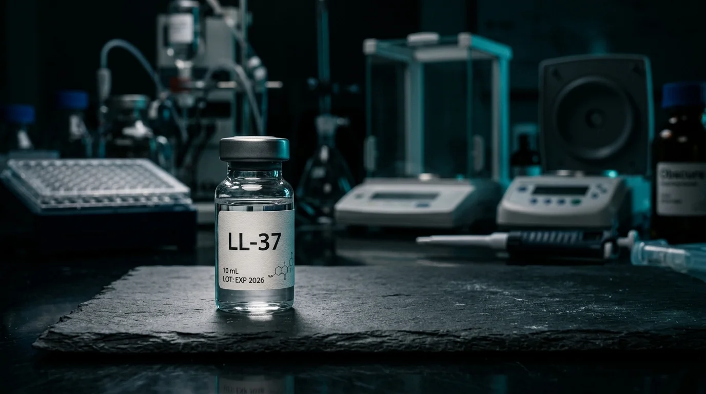
⚠️ Research chemical. Not for human administration. All data referenced is from preclinical studies (in vitro / animal models) unless otherwise noted.
⏱️ Reading time: ~16 min | Updated: May 22, 2026
LL-37: The Human Cathelicidin That Rewires Wound Healing and Antimicrobial Defense
Chronic wounds kill people. Not from infection alone — from the silent absence of a single peptide. When Heilborn and colleagues biopsied the edge of acute healing wounds versus non-healing chronic ulcers, they found something stark: LL-37 was everywhere in the healing tissue, and essentially gone from the chronic wound epithelium (PMID 12603850). That 2003 paper in Journal of Investigative Dermatology quietly shifted how the field understood why some wounds refuse to close.
LL-37 is the only human cathelicidin — a 37-amino acid host defense peptide that sits at the intersection of antimicrobial killing, wound repair, and immune modulation. What makes the research genuinely interesting isn't any single function. It's the fact that the same peptide that disrupts bacterial membranes also recruits neutrophils, accelerates keratinocyte migration, and — in the wrong proteolytic context — triggers the inflammatory cascades driving rosacea. Understanding the data means understanding the context.
Molecular Profile & Structure
LL-37 is a 37-residue cationic peptide with the sequence LLGDFFRKSKEKIGKEFKRIVQRIKDFLRNLVPRTES. The name derives from its two N-terminal leucines (LL) and its 37-amino-acid length. Key properties:
- CAS number: 154947-66-7
- Molecular weight: ~4,493 Da
- Net charge: +6 at physiological pH
- Secondary structure: predominantly alpha-helical in membrane-mimicking environments
- Amphipathicity: hydrophobic and cationic faces segregate when helical — critical for membrane activity
- Gene: CAMP gene, chromosome 3p21.3
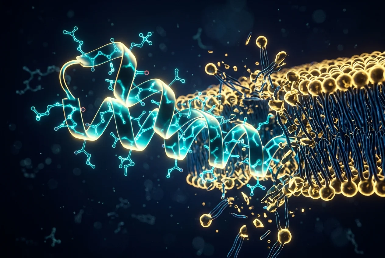
The amphipathic alpha-helix is the structural engine of its antimicrobial activity. In aqueous solution, LL-37 is largely unstructured. Contact with bacterial membranes or membrane-mimicking lipid environments induces rapid helical folding — a structural transition that makes the peptide insert itself into and disrupt lipid bilayers. Zasloff's landmark 2002 Nature review placed this mechanism within the broader framework of host defense peptides across multicellular organisms (PMID 11807545).
What separates LL-37 from many antimicrobial peptides: its cationic charge allows preferential electrostatic attraction to bacterial membranes (which carry net negative charge via phosphatidylglycerol and lipopolysaccharide), while the zwitterionic outer leaflet of mammalian cell membranes provides relative selectivity. "Relative" being operative — cytotoxicity at high concentrations is a known limitation, discussed in the limitations section.
Biosynthesis: From hCAP18 to LL-37
LL-37 isn't stored or secreted directly. It is proteolytically processed from its 18 kDa precursor, hCAP18 (human Cationic Antimicrobial Protein, 18 kDa). The processing enzymes differ by tissue:
- Neutrophils: serine protease 3 (PR3) cleaves hCAP18 to release LL-37 from specific granules during degranulation
- Keratinocytes: kallikrein-5 (KLK5) and kallikrein-7 (KLK7) generate LL-37 and shorter fragments — critically, the balance of kallikrein isoforms determines which fragments are produced
- Plasma: gastricsin-like enzymes in the male reproductive tract generate a slightly different fragment (ALL-38)
The kallikrein-5/LL-37 axis in skin is particularly important. Normal keratinocyte proteolytic processing produces LL-37. But in rosacea-affected skin, altered KLK5 activity generates aberrant processing products that trigger TLR2-mediated inflammation. Same precursor, different products, opposite biological outcomes. This is why the rosacea story is genuinely complex — and why blocking LL-37 globally would be the wrong therapeutic strategy.
Expression is inducible. Baseline LL-37/hCAP18 levels are low in healthy skin. Bacterial products (LPS, lipoteichoic acid), inflammatory cytokines (IL-1β, TNF-α), vitamin D, and physical wounding all strongly upregulate CAMP gene expression. This inducibility is a feature, not a bug — it allows tissue-specific, context-dependent deployment of host defense.
Antimicrobial Mechanism: How It Kills
There are three proposed models for LL-37 membrane disruption, and evidence supports elements of all three depending on peptide concentration and target membrane composition:
- Barrel-stave model: helical peptides insert perpendicularly to form a transmembrane barrel pore — relevant at high local concentrations
- Carpet model: peptides coat the membrane surface like a carpet, reaching a threshold density that disrupts membrane integrity and causes disintegration — most relevant for LL-37 at broad antimicrobial concentrations
- Toroidal pore model: peptides and lipids together form a pore with continuous curvature — produces ion leakage, ATP release, and cell death
Biofilm disruption is an underappreciated part of LL-37's research profile. Standard antibiotics struggle against biofilms, in part because the extracellular matrix blocks penetration. LL-37 disrupts biofilm architecture directly — both by targeting the bacteria and by degrading biofilm matrix components. Pavelka et al. 2026 demonstrated that cathelicidins embedded in a polysaccharide-based hydrogel scaffold maintained antimicrobial and pro-healing activity against carbapenem-resistant Pseudomonas aeruginosa in a porcine wound model (PMID 42150643). That's clinically relevant because carbapenem-resistant organisms are among the hardest-to-treat wound pathogens.
Antimicrobial Spectrum
Preclinical data support activity against a broad range of organisms:
- Gram-positive bacteria: S. aureus (including MRSA strains in some models), S. epidermidis, Streptococcus pyogenes
- Gram-negative bacteria: P. aeruginosa, E. coli, K. pneumoniae
- Mycobacteria: activity against M. tuberculosis has been documented, though MICs are higher
- Fungi: Candida albicans, Cryptococcus species show susceptibility in vitro
- Enveloped viruses: membrane-disruption activity extends to lipid-enveloped viruses (herpes simplex, influenza) — though mechanism details differ
One thing to be clear about: effective antimicrobial concentrations of LL-37 (typically 1–8 µg/mL in vitro) don't translate simply to physiological concentrations achievable at wound sites. The proteolytic environment of infected wounds can degrade LL-37 rapidly — which is precisely why the 2025/2026 delivery system research (PLGA nanocarriers, hydrogel scaffolds) is so important as a field direction.
Wound Healing Research
The pivotal evidence comes from Heilborn JD et al., 2003 (PMID 12603850). Their biopsy study compared LL-37 expression across three conditions: normal skin, acute healing wounds (punch biopsies), and chronic venous leg ulcers. The findings were direct:
- Strong LL-37 expression at the leading edge of re-epithelializing acute wounds — specifically in keratinocytes migrating to cover the wound
- Expression was virtually absent in chronic ulcer epithelium — the tissue that had failed to re-epithelialize
- Ex vivo application of LL-37 to wound edge tissue promoted keratinocyte migration in a receptor-dependent manner
Mechanistically, LL-37 promotes wound closure through multiple pathways:
- Keratinocyte migration: LL-37 activates EGFR (Epidermal Growth Factor Receptor) — either directly or via transactivation — which drives the lamellipodia formation and cell migration required for re-epithelialization
- Angiogenesis: at sub-antimicrobial concentrations, LL-37 promotes endothelial cell migration and tube formation, supporting neovascularization of healing tissue
- Inflammation modulation: LL-37 is chemotactic for neutrophils, monocytes, and T cells via formyl peptide receptor-like 1 (FPRL1), orchestrating the cellular response to injury
- Anti-apoptotic signaling: evidence from in vitro models suggests LL-37 can protect keratinocytes from UV-induced and cytokine-induced apoptosis
The 2025 work by De Soricellis et al. extended this into applied delivery research: lipidized LL-37 loaded into PLGA nanocarriers demonstrated significantly enhanced wound healing in ex vivo skin models compared to free peptide (PMID 40316190). The lipidization modification and nanoencapsulation both serve to protect LL-37 from proteolytic degradation — a key obstacle in translating in vitro data to in vivo wound environments.
For researchers studying tissue repair peptides, LL-37's wound healing profile is distinct from BPC-157 and TB-500. While BPC-157 works primarily through VEGFR2/eNOS-mediated angiogenesis and structural repair, LL-37 specifically addresses the re-epithelialization phase and the antimicrobial burden simultaneously. In chronically infected wounds, that dual function is uniquely relevant. See our complete BPC-157 research guide for a detailed comparison of the angiogenic mechanisms.
Immunomodulation: The Dual Role
LL-37 doesn't fit the simple "anti-inflammatory" or "pro-inflammatory" category. It's context-dependent in a way that makes it biologically interesting and therapeutically complicated.
Anti-Endotoxin Activity
One of LL-37's most studied immunomodulatory properties is LPS neutralization. Lipopolysaccharide (LPS/endotoxin from Gram-negative bacteria) is a potent TLR4 activator — the driver of septic shock in bacterial infection. LL-37 binds and neutralizes LPS, preventing TLR4 activation and the downstream cytokine storm. At physiological concentrations in wound exudate, this could significantly modulate the local inflammatory response.
NF-κB Modulation
LL-37's relationship with NF-κB is bidirectional and concentration-dependent. At low concentrations in non-infected contexts, LL-37 can suppress NF-κB activation by sequestering LPS and other PAMPs before they engage PRRs. However, LL-37 itself — especially the aberrant fragments generated in rosacea — can activate NF-κB via TLR2 and NLRP3 inflammasome pathways. This distinguishes LL-37 sharply from KPV, which is a direct upstream IKK-β inhibitor with unambiguous NF-κB suppression. Compare these mechanisms in our KPV anti-inflammatory peptide guide.
Chemotaxis and Immune Cell Recruitment
Via FPRL1 (formyl peptide receptor-like 1, now called FPR2), LL-37 acts as a potent chemoattractant for:
• Neutrophils — the first-line responders to infection
• Monocytes and macrophages — critical for phagocytosis and wound resolution
• T cells and mast cells — linking innate to adaptive immunity
This chemotactic activity is relevant for understanding both LL-37's wound-healing benefit (appropriate immune orchestration) and its pathological role in rosacea (excessive mast cell recruitment).
LL-37 in Rosacea & Acne: When the Defender Becomes the Provocateur
The rosacea story is arguably the most fascinating aspect of LL-37 research — and one that fundamentally reframes how researchers think about host defense peptide dysregulation.
Skin biopsies from rosacea patients consistently show elevated hCAP18/LL-37 expression — but with abnormal processing. Rather than the standard 37-residue LL-37 peptide, the altered kallikrein-5 activity generates shorter fragments (including the 4-kDa peptide originally termed "RK-31") that potently activate TLR2. TLR2 activation in skin mast cells triggers:
- Mast cell degranulation → histamine and tryptase release → erythema and flushing
- VEGF upregulation → telangiectasia (visible blood vessels)
- Neutrophil recruitment → pustule formation in papulopustular rosacea
- MMP activation → structural skin remodeling in phymatous rosacea
The 2026 fractional microneedle radiofrequency study (Xu et al., PMID 41664378) used an LL-37-induced mouse model — essentially injecting LL-37 to create rosacea-like skin inflammation — as the standard preclinical benchmark for evaluating treatment interventions. That this model is now standard tells you how well-established the LL-37-rosacea connection is in the field.
Wang & Zhou's 2026 review (PMID 41646975) places LL-37 within the broader rosacea pathogenesis framework, noting that the cathelicidin axis interacts with the microbiome (Demodex mites, Cutibacterium acnes), UV exposure, neurovascular dysregulation, and thermal triggers — all of which modulate KLK5 activity and therefore LL-37 fragment profiles.
Acne Research
In acne, the picture is different. Cutibacterium acnes (formerly P. acnes) induces LL-37 expression in keratinocytes via TLR2 signaling — this is part of the normal innate defense against the bacterium. LL-37 at physiological concentrations has direct antimicrobial activity against C. acnes. But simultaneously, LL-37 contributes to the inflammatory component of acne via cytokine induction. In atypical lesion profiles, this dual activity complicates interpretation. The data here are more preliminary than the rosacea literature.
LL-37 vs KPV vs BPC-157: Dermatological Research Comparison
When researchers study peptides for skin wound healing and dermatological applications, these three compounds come up repeatedly. Their mechanisms are entirely distinct — and understanding the differences matters for experiment design. This is UNIK LAB's cross-synthesis from the available preclinical literature.
↔ Scroll horizontally to view the full table on mobile.
| Parameter | LL-37 | KPV | BPC-157 |
|---|---|---|---|
| Origin | Human cathelicidin — processed from hCAP18 (innate immune system) | Synthetic fragment of α-MSH C-terminus (melanocortin system) | Synthetic pentadecapeptide derived from gastric juice protein BPC |
| Size | 37 aa / ~4,493 Da | 3 aa / ~314 Da | 15 aa / ~1,419 Da |
| Primary mechanism | Membrane disruption (carpet/toroidal model) + EGFR/FPRL1 signaling | NF-κB inhibition via IKK-β suppression; PepT1-mediated intracellular delivery | VEGFR2/eNOS upregulation; angiogenesis, collagen synthesis, tendon repair |
| Antimicrobial activity | Strong (broad spectrum, biofilm-active, MIC 1–8 µg/mL) | None documented | None documented |
| Wound healing evidence | Strong — re-epithelialization, keratinocyte migration, EGFR activation (PMID 12603850) | Moderate — skin inflammation models, epithelial barrier support | Strong — angiogenesis, tendon/muscle, broad tissue repair models |
| Rosacea/acne relevance | Central — dysregulated LL-37 fragments drive rosacea pathogenesis | Moderate — anti-inflammatory in skin keratinocyte models | Minimal — no published rosacea/acne-specific data |
| Effect size in preclinical models | Large for antimicrobial; moderate–large for wound re-epithelialization | Large for colitis; moderate for skin inflammation | Large for tendon/muscle repair; moderate for skin wound closure |
| Key limitation | Rapid proteolysis in wound environment; cytotoxicity at high concentrations; dual inflammatory role | Limited in vivo skin data; mostly intestinal models | Mechanism not fully elucidated; no human RCT data |
| Evidence level (2026) | Preclinical + some early Phase I data (infection/wound) | Preclinical only | Preclinical only |
| Delivery challenge | High — requires nanoencapsulation or lipidization to resist proteolysis | Low — oral bioavailability via PepT1 transporter | Moderate — stable in solution, subcutaneous or oral in rodent models |
Delivery, Stability & Formulation Research
LL-37's biggest translational obstacle isn't biological activity — the in vitro data are robust. It's delivery. The proteolytic environment of infected wounds, the short serum half-life, and concentration-dependent cytotoxicity all create formulation challenges that active research is addressing.
PLGA Nanocarriers
De Soricellis et al. 2025 (PMID 40316190) developed lipidized LL-37 in poly(lactic-co-glycolic acid) nanocarriers. Lipidization increases membrane affinity and reduces proteolytic access to the peptide backbone. The PLGA encapsulation provides sustained release over 48–72 hours. Ex vivo skin wound models showed significantly accelerated wound closure compared to free LL-37. This approach effectively addresses the half-life problem at the cost of formulation complexity.
Hydrogel Scaffolds
Pavelka et al. 2026 (PMID 42150643) used a polysaccharide-based hydrogel to embed cathelicidins for treatment of carbapenem-resistant Pseudomonas aeruginosa wound infections. The hydrogel scaffold serves dual purposes: physical wound coverage and sustained local peptide release. The porcine model is considered highly predictive of human wound healing due to similar skin architecture and wound contraction dynamics. The combination of pro-healing and antimicrobial activity within a single formulation represents the direction of applied LL-37 research.
Research-Grade LL-37: What to Look For
For laboratory use, research-grade LL-37 should meet:
- ≥98% purity by HPLC
- Mass spectrometry confirmation of correct molecular weight (~4,493 Da)
- Sequence verification — 37 amino acids matching LLGDFFRKSKEKIGKEFKRIVQRIKDFLRNLVPRTES
- Endotoxin ≤1 EU/mg (LAL assay) for in vivo use
- Sterile filtered for cell-based assays
Before any laboratory use, review the Certificate of Analysis carefully — our guide on how to read a peptide CoA walks through exactly what to check. Reconstitution protocol for LL-37 follows standard practice: see our peptide reconstitution guide for sterile water vs. bacteriostatic water considerations and storage temperature recommendations.
Storage note: LL-37 in lyophilized form is stable at -20°C for extended periods. In solution, it should be used within 2–4 weeks when stored at 4°C with BSA carrier added to prevent adsorption to tube walls. Avoid repeated freeze-thaw cycles — each cycle can reduce activity by 15–20% in antimicrobial assays.
Research Limitations & Evidence Quality
Honest assessment: LL-37 has strong preclinical foundations but significant translational hurdles. What the evidence supports, and where it gets complicated:
What the Evidence Supports
- LL-37 deficiency correlates with impaired wound healing — the Heilborn 2003 biopsy data are solid and have been replicated
- Direct antimicrobial activity across a broad pathogen spectrum — well-documented in vitro across hundreds of publications
- Aberrant LL-37 fragments drive rosacea inflammation — mechanistically established, clinically supported by therapeutic response to treatments that normalize kallikrein activity
- Biofilm disruption — multiple in vitro and animal model demonstrations
Where It Gets Complicated
- Cytotoxicity window: the therapeutic window between antimicrobial and cytotoxic concentrations is narrow and highly model-dependent. Healthy mammalian cells are affected at concentrations 2–5× the bacterial MIC in many cell types
- Proteolytic degradation in vivo: half-life in wound fluid is measured in minutes, not hours. Delivery system research is the current bottleneck
- Dual immunological role: the same peptide protects in acute wounds and drives pathology in rosacea. Context matters at every level
- No phase III data: as of 2026, no large RCTs have been completed for any LL-37 formulation. Early-phase wound care trials exist but results are preliminary
- Salt sensitivity: LL-37's antimicrobial activity is strongly inhibited by physiological salt concentrations — meaning the MICs measured in low-salt buffers may not translate directly to wound fluid conditions
Sourcing Research-Grade LL-37
LL-37 is a complex 37-amino-acid peptide — significantly larger than tripeptides like KPV or even 15-residue peptides like BPC-157. Larger peptides have higher synthesis costs and more potential for sequence errors. Quality sourcing matters more, not less, at this size.
For researchers in Europe evaluating suppliers, see our curated overview of where to buy research peptides in 2026 — with specific quality criteria and CoA expectations. UNIK LAB's current peptide catalog is available via the research peptide shop.
Frequently Asked Questions
What is LL-37?
LL-37 is the sole human cathelicidin antimicrobial peptide, comprising 37 amino acids (CAS 154947-66-7). Processed from the precursor hCAP18 by serine protease 3 in neutrophils and by kallikreins in keratinocytes, it bridges antimicrobial defense, wound repair, and immune modulation. It is a research chemical not approved for human administration.
How does LL-37 kill bacteria?
Via electrostatic attraction to negatively charged bacterial membranes, followed by amphipathic alpha-helix insertion and disruption. This physical mechanism makes resistance development significantly harder than with conventional antibiotics. Preclinical MIC values range from 1–8 µg/mL against clinically relevant pathogens including MRSA and P. aeruginosa.
What does LL-37 do for wound healing?
Heilborn et al. 2003 (PMID 12603850) demonstrated that LL-37 is present at the leading edge of acute healing wounds and absent in chronic non-healing ulcer epithelium. Mechanistically, it promotes keratinocyte migration via EGFR activation, supports angiogenesis, and orchestrates immune cell recruitment — collectively driving the re-epithelialization phase of wound repair.
Is LL-37 involved in rosacea?
Yes — in rosacea, altered kallikrein-5 activity generates aberrant LL-37 fragments that activate TLR2, triggering mast cell degranulation, VEGF upregulation (telangiectasia), and neutrophil recruitment. This pro-inflammatory role is the opposite of LL-37's protective function in normal wound healing — same molecule, different processing, different biological outcomes.
How does LL-37 compare to KPV and BPC-157?
LL-37 is the only antimicrobially active compound of the three. For infected wound research, its dual antimicrobial + re-epithelialization profile is unique. KPV offers superior NF-κB pathway specificity. BPC-157 leads in angiogenic and structural repair models. All three are preclinical-only compounds as of 2026.
What are the main limitations of LL-37 research?
Rapid proteolytic degradation in wound environments (minutes-scale half-life), cytotoxicity at concentrations approaching antimicrobial activity, salt-sensitivity that reduces efficacy at physiological salt concentrations, and the dual immunological role complicating any therapeutic targeting approach. Active delivery research (PLGA nanocarriers, hydrogel scaffolds) is addressing the stability problem.
What purity standards apply to research-grade LL-37?
≥98% HPLC purity with mass spec confirmation of the 37-residue sequence and correct molecular weight (~4,493 Da). Endotoxin ≤1 EU/mg by LAL assay is mandatory for in vivo experiments. Review the full CoA before laboratory use — see our guide on reading peptide certificates of analysis.
References
- Heilborn JD et al., J Invest Dermatol 2003;120(3):379-89. PMID 12603850
- Zasloff M. Nature 2002;415(6870):389-95. PMID 11807545
- De Soricellis C et al., Int J Pharm 2025;677:125668. PMID 40316190
- Xu Z et al., J Cosmet Dermatol 2026;25(2):e70727. PMID 41664378
- Wang H, Zhou C. Front Immunol 2026;16:1705588. PMID 41646975
- Pavelka A et al., Int J Pharm 2026. PMID 42150643
Available from UNIK LAB
Research Peptides
Browse UNIK LAB's research-grade peptide catalog — HPLC verified ≥98% · CoA included · EU dispatch